香港神經肌肉疾病學會

Putting Brains in Muscles

Towards an Accessible Web-Based Health Education Platform on Neuromuscular Diseases

Muscle Magnetic Resonance Imaging

What is Muscle Magnetic Resonance Imaging?

Muscle Magnetic Resonance Imaging (MRI) plays significant role in the diagnosis of neuromuscular disorders. It can not only precisely reveal the changes of muscle morphology and volume, but also assess the muscle fatty atrophy and edematous extent. It has far more precise localization competence compared with traditional neurological examination or electromyogram (EMG). Furthermore, some genetic neuromuscular disorders have unique and characteristic MRI muscle pattern of fatty atrophic and edematous changes, enabling its perfect use in localization, characterization, and even direct diagnosis. Muscle MRI is currently applied in precise localization before EMG or muscle biopsy, guiding further genetic testing targets, and evaluate the functional status and prognosis of patients. It is recommended that all the patients with suspected neuromuscular disorders undergo muscle MRI, potentiating the diagnostic accuracy.

Case 1: collagen VI-related myopathy (Bethlem myopathy type 1)

The thigh muscle MRI showed characteristic “target and sandwich sign”. 

Case 2: GNE myopathy

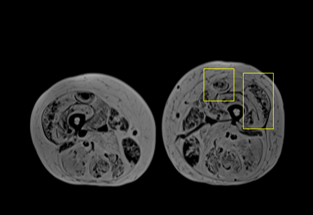
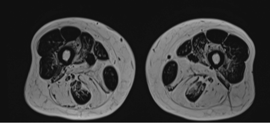
Thigh muscle showed confined fatty atrophy involving rectus femoris and hamstring muscles.

Case 3: Myofibrillar myopathy type 5

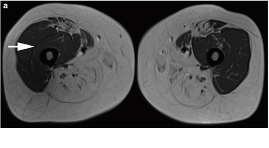
The thigh muscle MRI revealed fatty atrophy involving hamstring muscles, adductors, and vastus intermediate. Interestingly, the semitendinosus is relatively spared.

The Hong Kong Society of Neuromuscular Diseases

Search
Close this search box.